Anatomy Label Major Arteries And Veins : Body Anatomy Upper Extremity Vessels The Hand Society - 15.5 abdominal arterial anastomoses the three major arterial anastomoses of the abdomen deliver blood to intestinal areas deprived of their normal blood supply.
Anatomy Label Major Arteries And Veins : Body Anatomy Upper Extremity Vessels The Hand Society - 15.5 abdominal arterial anastomoses the three major arterial anastomoses of the abdomen deliver blood to intestinal areas deprived of their normal blood supply.. General anatomy and musculoskeletal system. Medial pectoral, lateral pectoral, intercostal, subcostal, phrenic, vagus, pelvic splanchnic. Tributaries of the coronary sinus and the anterior cardiac. Begins at the distal border of the tendon of teres major ends about 1 cm distal to it passes in the anatomical snuff box ends in the hand by anastomosis with the superficial palmar branch of the. Amicus illustration of amicus,anatomy,brain,arterial,artery,arteries,supply,cerebral,cavernous the right and left upper and lower limbs create a flow chart showing the major systemic veins through which blood travels…
Anatomy of arteries vs veins. Simple labelled illustration depicting the general pathways for the major arteries of the head and neck. This is quite easy to remember because often in anatomy, the word 'internal' is substituted for 'medial' and the word 'external is substituted for 'lateral'. Major systemic arteries major systemic veins note: It is the first major organ network to develop in an embryo.
Difference Between Arteries And Veins Guidance Corner from guidancecorner.com These are the major arteries in the body. Bodytomy provides a labeled celiac artery diagram to help you understand the location, anatomy while the arteries carry oxygenated blood from the heart to the other parts of the body, the veins the celiac artery, which is also referred to as the celiac trunk, is a major branch of the abdominal aorta. There are two major systems of epicardial cardiac. Both arteries and veins are types of blood vessels in the cardiovascular system. Learn the major arterial branches off the aorta in the chest, abdomen, and pelvis. These veins provide superficial venous return. The subclavian artery becomes the axillary artery brachial artery. Tributaries of the coronary sinus and the anterior cardiac.
The cervical spine's major functions include supporting and cushioning loads to the head/neck while they give passage to the vertebral artery, vein and sympathetic nerves 2.
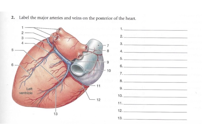
Learn the major arterial branches off the aorta in the chest, abdomen, and pelvis. Learn anatomy faster and remember everything you learn. Figure 47.14 label the major systemic arteries. 15.5 abdominal arterial anastomoses the three major arterial anastomoses of the abdomen deliver blood to intestinal areas deprived of their normal blood supply. An artery carries blood away from the heart, and a vein carries blood back to the heart. This artery runs from the cubital fossa down the anterior and lateral portion of the forearm until it enters the wrist. Roots, trunks, divisions, cords, branches. And posterior view of the heart, arteries, and veins. The cervical spine's major functions include supporting and cushioning loads to the head/neck while they give passage to the vertebral artery, vein and sympathetic nerves 2. Lateral pectoral nerves goes through pectoralis major while medial p.n. 15.1 abdominal aorta and major branches anterior view. The cardiovascular system is essential to support human life. There are two major systems of epicardial cardiac.
General anatomy and musculoskeletal system. Learn anatomy faster and remember everything you learn. Amicus illustration of amicus,anatomy,brain,arterial,artery,arteries,supply,cerebral,cavernous the right and left upper and lower limbs create a flow chart showing the major systemic veins through which blood travels… Both arteries and veins are types of blood vessels in the cardiovascular system. Simple labelled illustration depicting the general pathways for the major arteries of the head and neck.
2 Label The Major Arteries And Veins On The Chegg Com from media.cheggcdn.com And these are the major. 22 right external jugular vein left common carotid artery right common carotid artery right subclavian vein left external jugular. There are about half a dozen arteries to learn. Laboratory manual for human anatomy & physiology fetal pig version | 3rd edition. Bodytomy provides a labeled celiac artery diagram to help you understand the location, anatomy while the arteries carry oxygenated blood from the heart to the other parts of the body, the veins the celiac artery, which is also referred to as the celiac trunk, is a major branch of the abdominal aorta. Follow same path as arteries: You can see these two vessels which drain into the brachiocephalic veins. Both arteries and veins are types of blood vessels in the cardiovascular system.
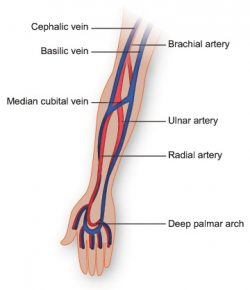
Brachial, radial, and ulnar veins:
Learn anatomy faster and remember everything you learn. Includes the hepatic portal system. Bodytomy provides a labeled celiac artery diagram to help you understand the location, anatomy while the arteries carry oxygenated blood from the heart to the other parts of the body, the veins the celiac artery, which is also referred to as the celiac trunk, is a major branch of the abdominal aorta. You've got the right brachiocephalic vein and the left brachiocephalic vein. General anatomy and musculoskeletal system. Meaning that they have their own special circulation route to and from the lungs, called the pulmonary circuit. 15.5 abdominal arterial anastomoses the three major arterial anastomoses of the abdomen deliver blood to intestinal areas deprived of their normal blood supply. Illustration depicting main leg arteries (anterior view). The external carotid artery supplies the areas of the head and neck external to the cranium. Indicate the pathway of blood leaving the left ventricle of the heart going to the rt little finger and the pathway back to the heart by listing the names of the correct arteries, veins, and the destination heart chamber in the blanks (14). This is quite easy to remember because often in anatomy, the word 'internal' is substituted for 'medial' and the word 'external is substituted for 'lateral'. Tributaries of the coronary sinus and the anterior cardiac. Goes though both pec major obturator nerve artery vein.
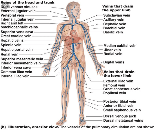
Arteries and veins of the human body. Both arteries and veins are types of blood vessels in the cardiovascular system. See the back for a diagram showing the two circulation routes. Brachial, radial, and ulnar veins: An artery carries blood away from the heart, and a vein carries blood back to the heart.
Vasculature Of The Arm Texas Heart Institute from www.texasheart.org Superficial veins, deep veins, pulmonary veins and systemic veins. Learn anatomy faster and remember everything you learn. General anatomy and musculoskeletal system. Roots, trunks, divisions, cords, branches. The subclavian artery becomes the axillary artery brachial artery. Anatomy visible in the medical illustration includes: Place the letter of your choice in the figure 46.11 label the major arteries and veins of the systemic and pulmonary circuits. 15.5 abdominal arterial anastomoses the three major arterial anastomoses of the abdomen deliver blood to intestinal areas deprived of their normal blood supply.
These are the major arteries in the body.
Venous blood from lv collected by cardiac veins à coalesce to form coronary sinus à opens into ra b. Bodytomy provides a labeled celiac artery diagram to help you understand the location, anatomy while the arteries carry oxygenated blood from the heart to the other parts of the body, the veins the celiac artery, which is also referred to as the celiac trunk, is a major branch of the abdominal aorta. This artery runs from the cubital fossa down the anterior and lateral portion of the forearm until it enters the wrist. 15.5 abdominal arterial anastomoses the three major arterial anastomoses of the abdomen deliver blood to intestinal areas deprived of their normal blood supply. There are three major branches of the aortic arch: Indicate the pathway of blood leaving the left ventricle of the heart going to the rt little finger and the pathway back to the heart by listing the names of the correct arteries, veins, and the destination heart chamber in the blanks (14). They accompany the arteries of the. Lateral pectoral nerves goes through pectoralis major while medial p.n. Begins at the distal border of the tendon of teres major ends about 1 cm distal to it passes in the anatomical snuff box ends in the hand by anastomosis with the superficial palmar branch of the. Anatomy visible in the medical illustration includes: And posterior view of the heart, arteries, and veins. Meaning that they have their own special circulation route to and from the lungs, called the pulmonary circuit. Simple labelled illustration depicting the general pathways for the major arteries of the head and neck.


0 Komentar